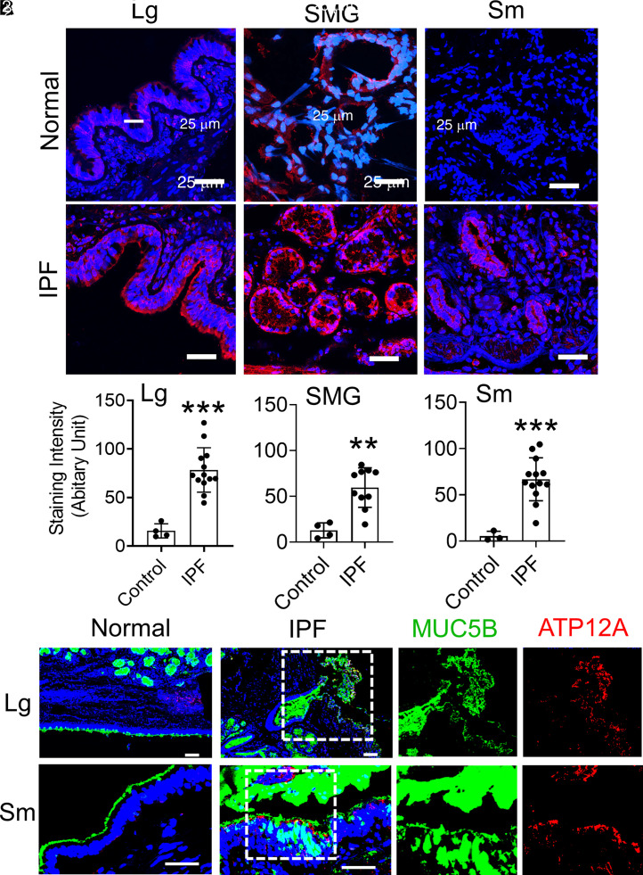
Figure 1.
ATP12A (adenovirus-expressing mouse ATP12A [Ad-ATP12A]) and MUC5B (Mucin 5B) protein expression in human lung explants. (A) Representative confocal microscope images showing immunodetection of ATP12A (red) by immunofluorescence. Nuclei were counterstained by DAPI (blue). Scale bars, 25 μm. Images show the large airways (Lg), SMG, and small airways (Sm) of normal human lungs (upper panel) and human lungs with idiopathic pulmonary fibrosis (IPF) (lower panel). Sm are defined as airways having a diameter that is less than 2 mm. ATP12A overexpression was found in large and Sm as well as in the submucosal glands of IPF. (B) ATP12A immunofluorescence staining intensity quantification charts. Data are expressed as mean ± SD of 4 normal and 13 IPF lung samples. At least six lung sections were examined per donor, and ATP12A expression intensity was quantified in more than six Sm per donor. **P < 0.01 and ***P < 0.001, compared with control, respectively. (C) Representative confocal microscope images showing immunodetection of ATP12A (red) and MUC5B (green) in Lg and Sm of normal and IPF human lungs. Nuclei were counterstained by DAPI (blue). Scale bars, 25 μm. ATP12A and MUC5B were overexpressed in both Lg and Sm of IPF lungs. The dotted line squares indicate the area in the section that have been magnified to show the co-expression of MUC5B and ATP12A. SMG = submucosal glands.

