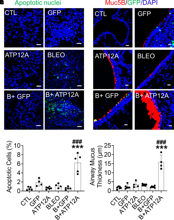
Figure 3.
Viral vector–mediated ATP12A expression in mouse airways worsens BLEO-induced alveolar epithelium apoptosis and airway mucus accumulation. (A) Confocal microscope images show cellular apoptosis of lung epithelial cells by TUNEL staining. Apoptotic cell nuclei are stained green. Scale bars, 25 μm. (B) The chart shows the percentage of apoptotic cells in mouse lungs. Data are expressed as mean ± SD, with n ⩾ 5 animals per group. (C) Confocal microscope images show immunodetection of MUC5B (red) by immunofluorescence. Nuclei were counterstained by DAPI (blue). Scale bars, 25 μm. (D) The chart shows airway mucus thickness in mouse lungs. Data are expressed as mean ± SD. n ⩾ 5 animals per group. ***P < 0.001, compared with CTL, respectively. ###P < 0.001, compared with BLEO-treated group.

