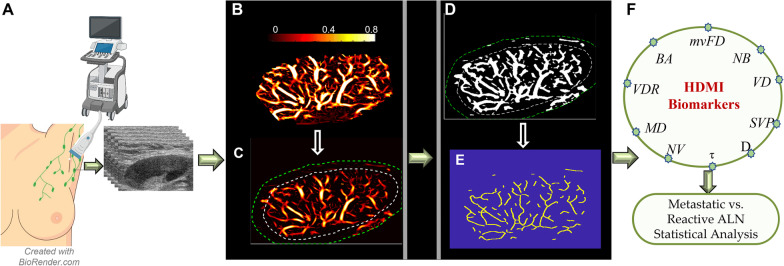
Fig. 1.
HDMI image acquisition and a set of processing for image segmentation to extract vessel biomarkers: A HDMI image acquisition of ALN, B Microvasculature image of a metastatic ALN, C Defined ROI with 2 mm dilation of the ALN, D Conversion of the microvasculature image into a binary image, E skeleton image of vessel network, F HDMI biomarkers extracted for the skeleton to be used for distinguishing the metastatic ALN from reactive. HDMI: High definition microvessel imaging; ALN: Axillary lymph node; mvFD: Microvessel fractal dimension; NB: Number of branch points; VD: Vessel density; SVP: Spatial vascularity pattern; D: Diameter; τ: Tortuosity; NV: Number of vessel segments; MD: Murray’s deviation; VDR: Vessel density ratio; BA: Bifurcation angle.

