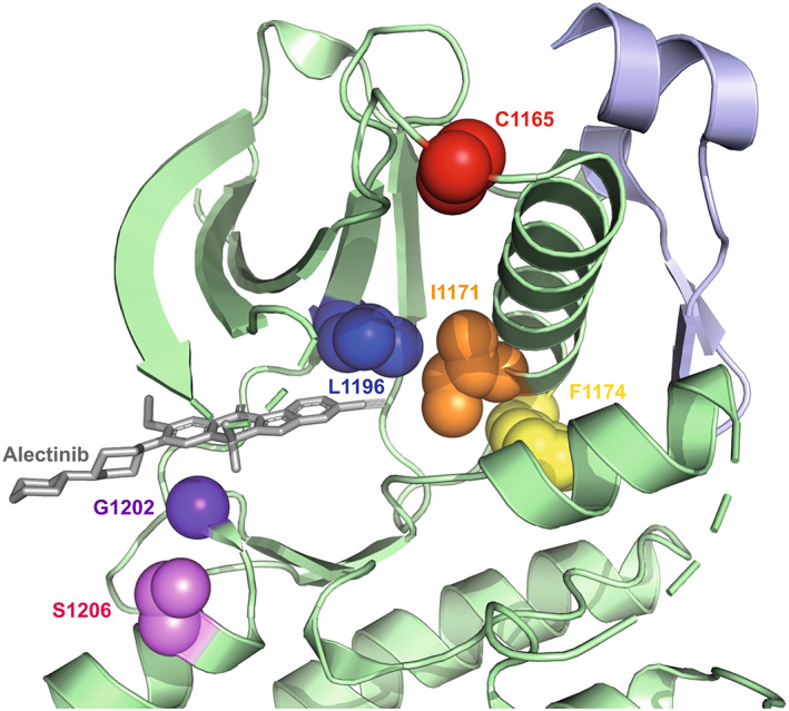
Fig. 2.

Structure of ALK kinase domain and key mutation sites. View of ALK kinase domain (green) bound to alectinib (grey) centred on the active site highlighting the key residues that are frequently mutated mapped onto the crystal structure (PDB:3AOX). Red: C1156 residue, Orange: I1171 residue, Yellow: F1174 residue, Blue: L1196 residue, Indigo: G1202 residue, Violet: S1206 residue. The juxtamembrane region of ALK is shown in light purple.
