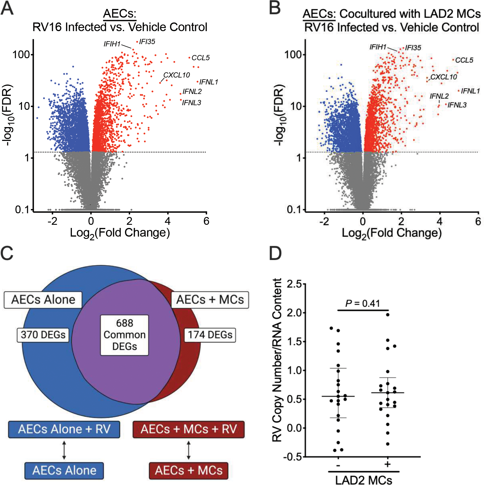
Figure 2.

Mast cells (MCs) modestly alter airway epithelial cell (AEC) responses to human rhinovirus A16 (RV16) infection. (A, B) Volcano plots showing fold-change differences between RV16-infected AECs and AECs treated with vehicle control in the absence of LAD2 MCs (A) or presence of LAD2 MCs (B). (C) Venn diagram demonstrating overlapping and non-overlapping differentially expressed genes (FDR <0.05 and a log2 fold change ≥1.0 or ≤−1.0) between AECs at baseline and following RV16 infection in the presence or absence of MCs. (D) RV16 copy numbers relative to RNA concentration between AECs cultured in the presence or absence of MCs. RV copy numbers are log-transformed. P value represents the results from a Wilcoxon matched-pairs signed rank test.
