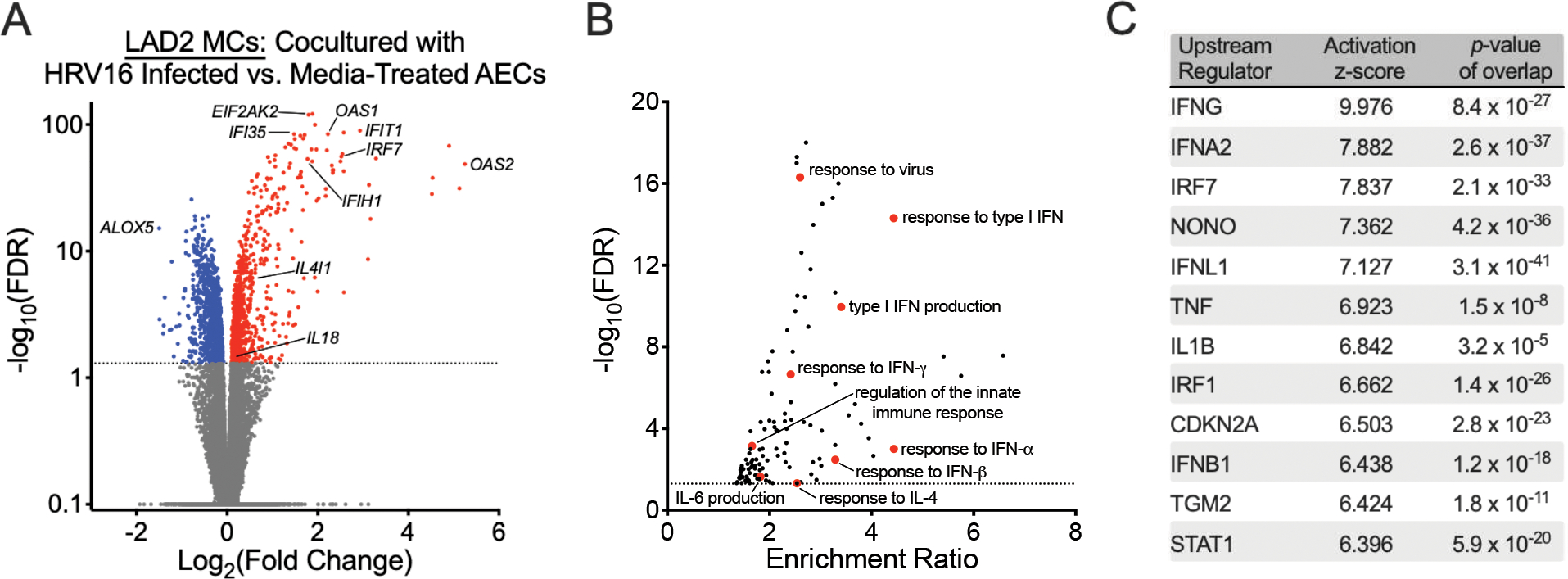
Figure 3.

Human rhinovirus A16 (RV16) infection of airway epithelial cells (AECs) alter mast cell (MC) gene expression patterns. (A) A volcano plot showing fold-change differences in gene expression between LAD2 MCs cultured in the presence of RV16-infected AECs versus AECs treated with vehicle control identified using a paired analysis. Red indicates genes that have significantly higher expression and blue indicates genes that have significantly lower expression in the LAD2 MCs cultured in the presence of RV16-infected AEC group (FDR <0.05). (B) Gene Ontology (GO) biologic processes overrepresented amongst the 1897 differentially expressed genes between AECs cultured in the presence of RV16-infected AECs versus AECs treated with vehicle control using WebGestalt 2019 (FDR <0.05). (C) Upstream regulator analysis of the 1897 differentially expressed genes between AECs cultured in the presence of RV16-infected AECs versus AECs treated with vehicle control using Ingenuity Pathway Analysis.
