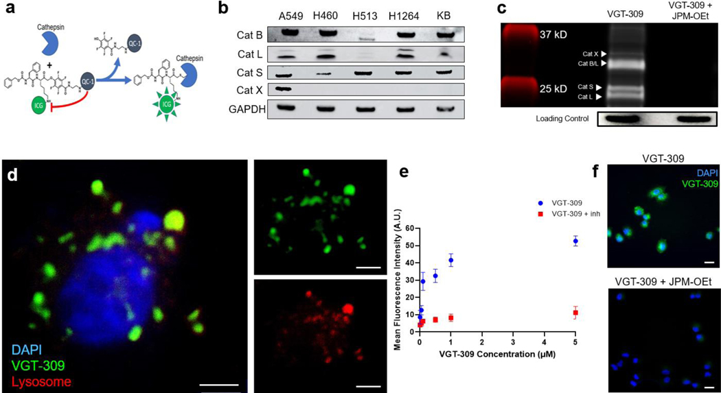
Figure 1.
VGT-309 labels human lung cancer cell lines in a cathepsin activity-dependent manner. a. Mechanism of action of VGT-309. The ICG fluorophore is quenched by the QC-1 quencher until it is cleaved upon covalent binding by cysteine cathepsins. b. Western blot analysis of cathepsin expression in human non-small cell lung cancer cell lines with KB (human cervical carcinoma) used as a positive control. c. SDS-PAGE analysis of probe-labeled species in H1264 (human pulmonary squamous cell carcinoma) cells with and without pretreatment with 100 uM JPM-OEt, a broad spectrum cathepsin inhibitor. d. Fluorescence microscopy of A549 (human pulmonary adenocarcinoma) cells at 100x magnification 1 hour after treatment with 1 uM VGT-309. Cells were co-stained with DAPI and Lyso-Tracker. VGT-309 labeling (shown in green) is predominantly lysosomal (shown in red) in distribution. Overlay images are shown at left with VGT-309 channel and Lyso-Tracker channels shown at right. Scale bars represent 5 μm. e. Fluorescence intensity of A549 cells 1 hour after administration of increasing concentrations of VGT-309. Cells were pretreated for 30 minutes with either JPM-OEt or DMSO vehicle prior to VGT-309 administration. f. Fluorescence microscopy showing VGT-309 fluorescence in A549 cells with and without pretreatment with JPM-OEt, showing cathepsin-activity dependence of fluorescence. Scale bars represent 20 μm.

