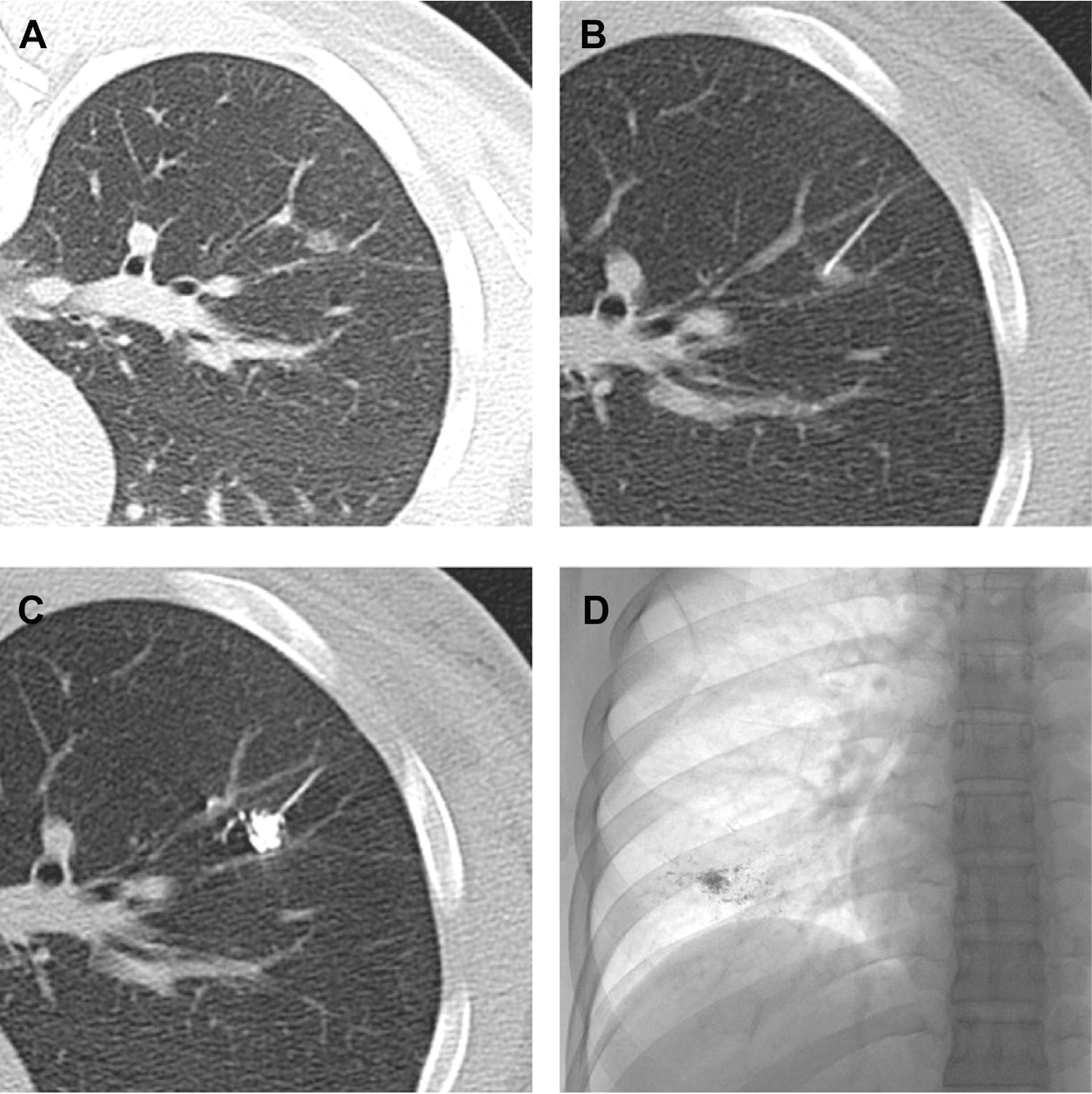
Fig. 3.

Computed tomographic fluoroscopic guided lipiodol-marking procedure for ground-glass opaque (GGO) nodules. (A) A small GGO in the right lower lobe. (B) A percutaneous transhepatic cholangiography needle (23 gauge) is inserted in the center of the GGO nodule. (C) Lipiodol 0.3 mL is then injected into the nodule. (D) A radiopaque spot is clearly demonstrated on the plain chest radiograph. (Obtained from Thoracic Surgery Clinics. Elsevier.)
