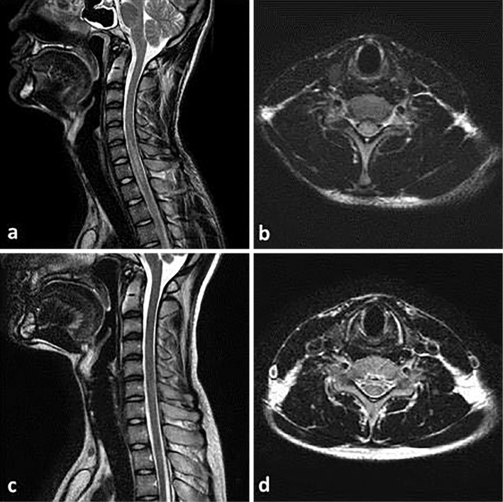
Figure 1.
Images of cervical spine MRI. (a) Pre-treatment T2-weighted sagittal images of the cervical spine reveal longitudinally extensive transverse myelitis. (b) Pre-treatment T2-weighted axial images of the cervical spine reveal transverse myelitis. (c) Post-treatment T2-weighted sagittal images of the cervical spine reveal the disappearance of myelitis. (d) Post-treatment T2-weighted axial images of the cervical spine reveal the disappearance of myelitis.

