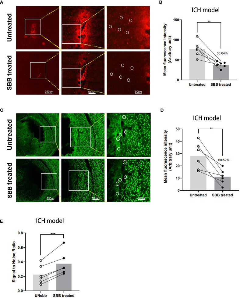
Figure 3.
Comparison of the IL-10 and NeuN fluorescence signal of brain tissues damaged by intracerebral hemorrhage before and after Sudan black B (SBB) treatment. (A) Striatal injection of Cy5.5-labeled IL-10 was used to evaluate the fluorescence of intracerebral hemorrhage-damaged brain tissues in the same brain sections, before and after SBB treatment, under a fluorescence microscope with the Tx Red filter. The white circles indicate representative regions of the significant difference in fluorescence signal before and after SBB treatment. (B) SBB treatment eliminated tissue autofluorescence by 50.64% of the untreated level (**p=0.0029; t=5.416; paired t-test; n=6 mice/group). (C) Anti-NeuN antibody labeled with Alexa Fluor 488 was used to assess the intensity of immunofluorescence of brain tissues in brain sections affected by ICH before and after SBB treatment under a fluorescence microscope using the FITC filter system. The white circles indicate representative regions of the significant difference in fluorescence signal before and after SBB treatment. (D) SBB treatment eliminated tissue autofluorescence by 60.52% of the untreated level (**p=0.0072; t=4.369; paired t-test; n=6 mice/group). (E) Compared to the untreated group, SBB treatment significantly improved the signal-to-noise ratio of fluorescence imaging (***p=0.0007; t=7.411; paired t-test; n=6 mice/group).

