Figure 2.
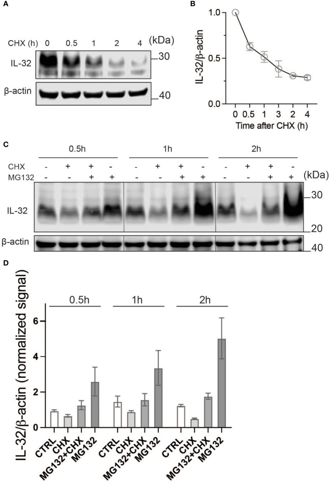
IL-32 has a high protein turnover. (A) JJN-3 cells were treated with CHX and harvested at the indicated time points. IL-32 protein levels were analyzed by WB. (B) Kinetics of IL-32 degradation in JJN3- cells. IL-32 protein signal intensity was quantified and normalized to loading control in n = 6 CHX chase experiments. The mean ±SEM is shown here. (C) JJN-3 cells were treated with 5 μg/ml CHX and 20 μM MG132 alone and in combination and harvested at the indicated time points. The figure shows one representative WB from n = 3 independent experiments. (D) Signal intensities of IL-32 and β-actin were quantified from n = 3 independent experiments performed as in (C), and the values from treated samples at each time point were normalized relative to the control sample.

