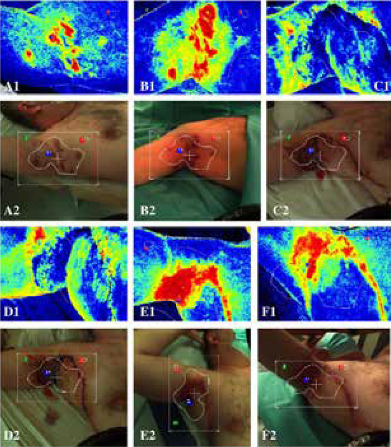
Figure 2.
A1–A2 – patient before surgical treatment – image taken in the outpatient clinic. B1–B2 – image taken before the operation (a progression of HS is seen – red colour), C1–C2 – image taken after the surgical procedure (co-graft of ADM and STSG and rotation flap) – pale STSG and pale rotation flap (ischemia) can be seen, D1–D2 – image taken 4 days after the operation, E1–E2 – image taken 2.5 months after surgical treatment (normal healing process), F1–F2 – image taken 3 months after surgical treatment (fully healed)

