
Figure 4.
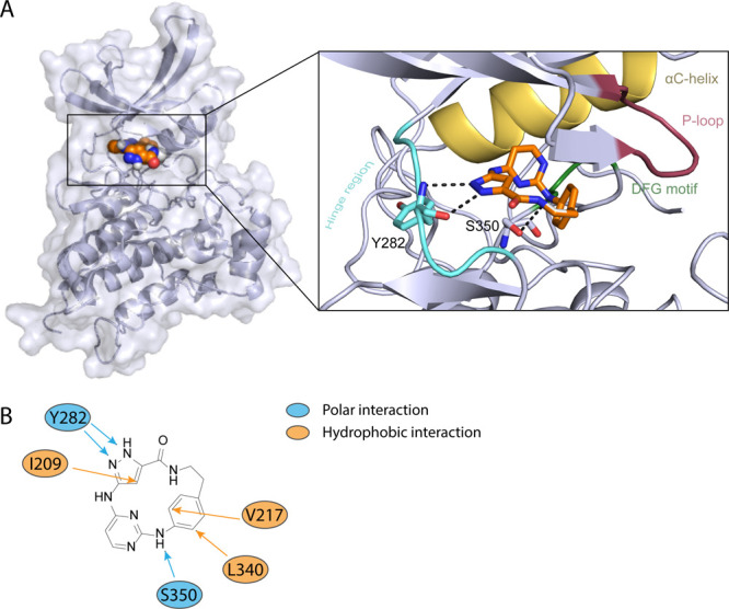
A. Binding mode of 8a with the kinase domain of BMPR2 determined by in silico docking. Macrocycle 8a was docked into the active conformation of BMPR2 (PDB: 6UNP). Hinge region is highlighted in light blue, P-loop red, DFG motif green, and αC-helix yellow, and the compound 8a is illustrated in orange. Docking poses were viewed by PyMOL, and protein–ligand interactions were analyzed using the PLIP.34 B. Interactions between the inhibitor and the amino acid residues of the protein kinase. Blue indicates a polar interaction and orange a hydrophobic interaction between the inhibitor 8a and BMPR2.