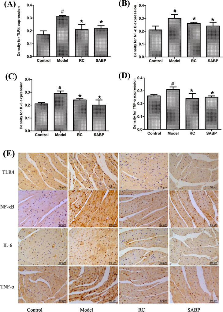
Fig. (5).
SABP suppressed TLR4, NF-κB, IL-6 and TNF-α expression in the heart. (A-D) Normalized quantitative data for TLR4, NF-κB, IL-6 and TNF-α protein expression levels; (E) The TLR4, NF-κB, IL-6 and TNF-α expression were displayed by immunohistochemistry staining. The arrows showed the staining for targeted protein. Values are expressed as mean ± SD, n = 4. #P < 0.05, compared with control group;*P < 0.05, compared with the model group.

