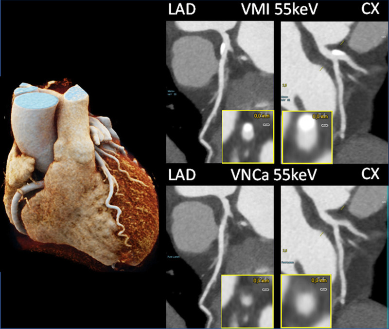
FIGURE 11.

Coronary CTA images of a patient with calcifications in the LAD and CX, scanned with a PCD-DSCT (120 kV, 144 × 0.4 mm, 0.25 seconds rotation time, 66 milliseconds temporal resolution). The calcifications shown in the 55 keV VMIs (top) are removed in the 55 keV virtual noncalcium images (bottom). Courtesy of Dr H. Alkadhi, University Hospital Zurich, Switzerland.
