
FIGURE 6.
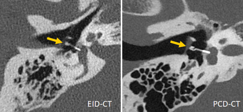
Inner ear scans acquired with a third-generation DSCT (left) and a first-generation PCD-DSCT, (right), demonstrating superior visualization of a Titanium K-piston stapes prosthesis (arrow, distal end diameter 0.4 mm) with PCD-CT. Courtesy of Dr A. van der Lugt, Erasmus Medical Center, Rotterdam, the Netherlands.