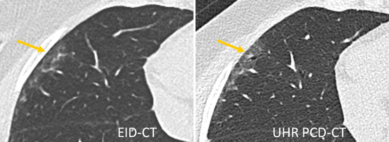
FIGURE 7.

Lung scan of a patient with interstitial lung disease acquired with a third-generation DSCT (left) and a first-generation PCD-DSCT, (right). The UHR PCD-CT scan confirms the presence of traction bronchiolectasis in the periphery of the right middle lobe (arrow), superimposed on mild ground-glass attenuation. This finding was missed on the EID-CT scan. Courtesy of Dr Martine Remy-Jardin, Hôpital Calmette, Lille, France.
