
FIGURE 8.
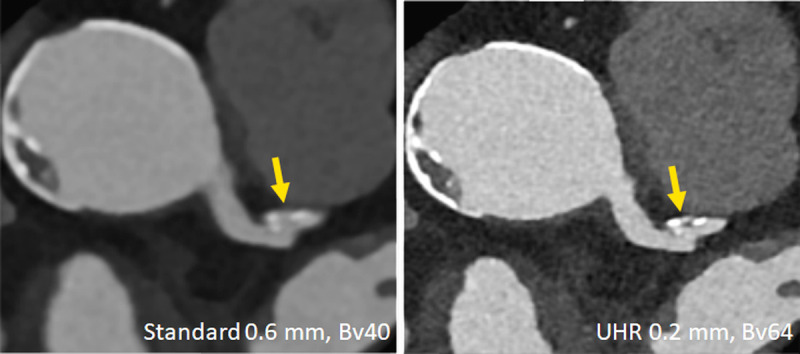
Clinical example demonstrating improved characterization of coronary plaques with UHR PCD-CT. A plaque that appears essentially calcified in a standard reconstruction (left) turns out to be mixed and smaller in volume in the UHR image (right). Image courtesy of Drs V. Mergen and H. Alkadhi, University Hospital Zurich, Switzerland.