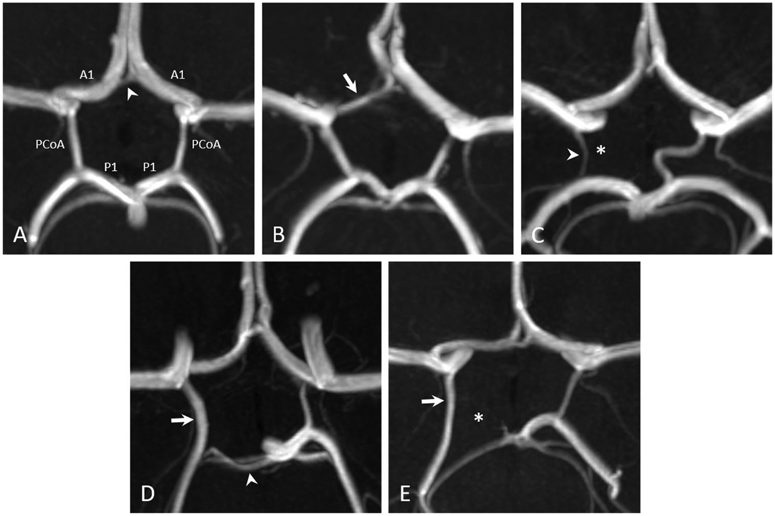
Fig. 1.

3D reconstructed time-of-flight magnetic resonance angiographic images of five different pediatric patients demonstrate (A) a complete circle of Willis (white arrowhead: anterior communicating artery), (B) a CoW with a hypoplastic right A1 segment (white arrow), (C) a CoW with an absent right posterior communicating artery (white asterisk; white arrowhead: right anterior choroidal artery), (D) a partial fetal-type right posterior cerebral artery (white arrow) with a hypoplastic right P1 segment (white arrowhead), and (E) a full fetal-type right posterior cerebral artery (white arrow) with an absent right P1 segment (white asterisk)
