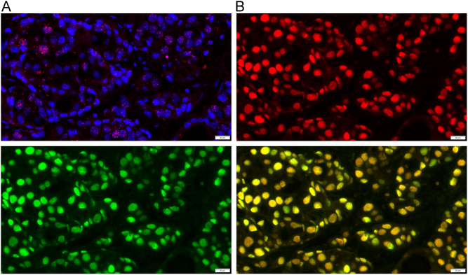
Figure 2.
Visualization of ERα homodimers and ERα immunoreactivity in formalin-fixed paraffin-embedded breast cancer tissues. (A) ERα homodimers is indicated by red dots. Nucleus is shown in blue (DAPI). (B) ERα immunoreactivity is shown in green and red, respectively. Double positive cells are shown in yellow.

 This work is licensed under a