. 2023 May 31;145(8):081002. doi: 10.1115/1.4062524

Fig. 6.

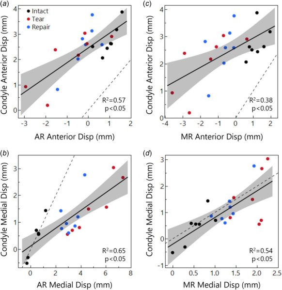
Displacement relationships between the medial condyle contact point and the meniscus AR (a and b) and MR (c and d), considering anterior (a and c) and medial (b and d) displacement components. The condyle moves further anteriorly than the meniscus (a and b), while medial displacements (b and d) are closer to a 1:1 relationship (dashed line) between the tissues. This 1:1 relationship is lost in the AR with tear and repair conditions because, with theanterior medial meniscus attachment released, the meniscus AR displaces far outside of the joint space with loading.

Displacement relationships between the medial condyle contact point and the meniscus AR (a and b) and MR (c and d), considering anterior (a and c) and medial (b and d) displacement components. The condyle moves further anteriorly than the meniscus (a and b), while medial displacements (b and d) are closer to a 1:1 relationship (dashed line) between the tissues. This 1:1 relationship is lost in the AR with tear and repair conditions because, with theanterior medial meniscus attachment released, the meniscus AR displaces far outside of the joint space with loading.