. 2023 May 15;12:e85410. doi: 10.7554/eLife.85410

Figure 3. R19G10 (PI-CA) neurons project to the corpora allata, which are required for summiting behavior.

(A) Left: Composite micrograph of dissected Aug21>GFP fly, showing GFP fluorescence in the corpora allata (CA) overlaid on bright field image. Right: Diagram of A with anatomical features labeled. PV = proventriculus. (B) Representative confocal micrograph of immunostained RC from an R19G10>syt-eGFP, DenMark fly. Synaptic terminals are visible as green puncta, including in the CA. Magenta is anti-JHAMT and marks the CA. Blue phalloidin counterstain marks actin. Labels as in A. (C) Summiting effect size estimate distribution of ablating the CA with diphtheria toxin (DTI). Effect size is calculated relative to effector-less sibling controls. (D) Representative micrographs of CA-ablated and effector-less, sibling, temperature-matched control flies (additional examples in Figure 3—figure supplement 1D). White arrows indicate the expected location of CA. e = eye, p = proboscis. (E) Summiting effect size estimate distributions of various concentrations of the CA-ablating drug precocene. Effect size is calculated relative to vehicle (acetone) control. For (C and E), effect sizes were estimated as in Figure 2; asterisks indicate statistically significant effects (*=p<0.05; **=p<0.01; ***p<0.001) by two-tailed t-test. Sample sizes of experimental and control experiments are given in black and gray, respectively.

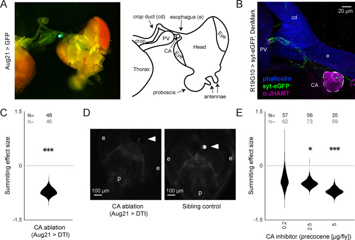
Figure 3.

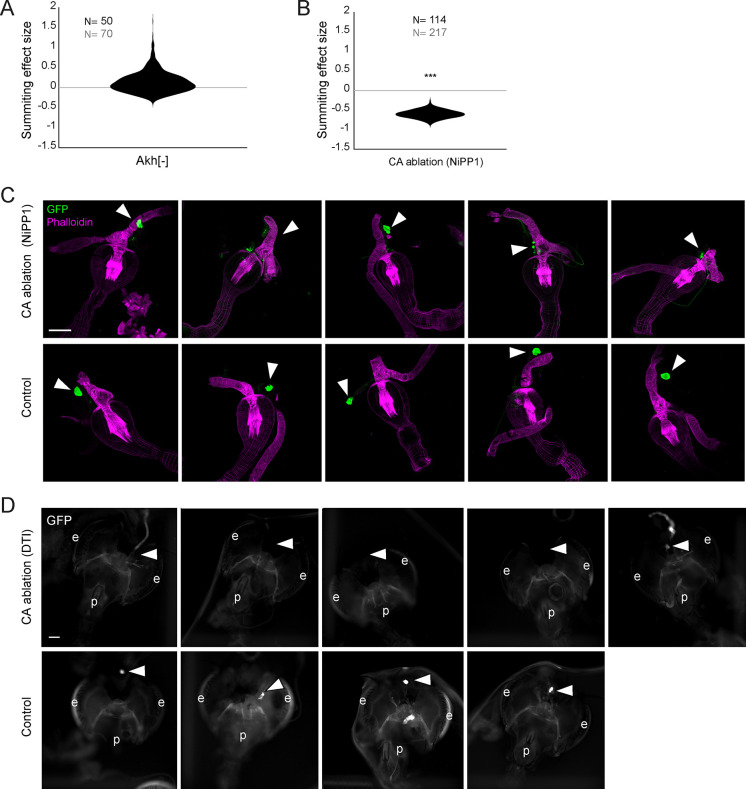
Figure 3—figure supplement 1. Supporting data for juvenile hormone involvement in summiting.

Figure 3—figure supplement 1.

(A) Summiting effect size estimate distribution for Akh- mutants, showing no effect compared to controls. Sample size of experimental animals is in black, and controls in gray. *=p<0.05; **=p<0.01; ***p<0.001 by two-tailed t-test. (B) As in (A) for the ablation of the corpora allata (CA) using NiPP1 effector (per Yamamoto et al., 2013). A significant reduction in summiting is seen compared to controls. (C) Confocal micrographs of the anterior foregut and retrocerebral complexes from Aug21>GFP, NiPP1 flies, and Aug21>GFP sibling controls. Green channel is GFP and magenta anti-actin phalloidin counterstain. Arrowheads indicate the observed or expected location of CA. Scale bar is 100 microns. CAs in Aug21>GFP, NiPP1 animals ranged from intact to absent. (D) Compound epifluorescence micrographs of the head, anterior foregut, and retrocerebral complexes of tub-Gal80, Aug21> GFP, DTI flies. In animals in which DTI expression was induced by thermal inactivation of the Gal80 repressor, only one sample showed potential residual CA; all other animals lacked CA.CA were observed in all controls. e = eye, p = proboscis.
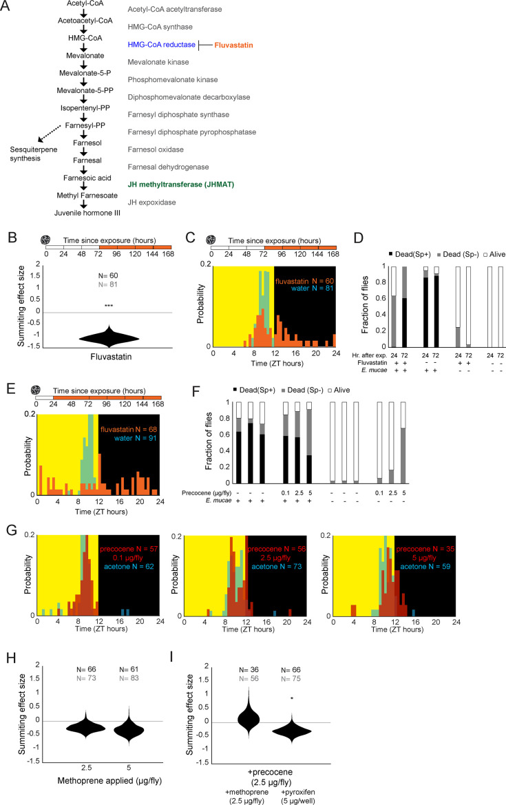
Figure 3—figure supplement 2. Additional experiments examining juvenile hormone involvement in summiting.

Figure 3—figure supplement 2.

(A) Synthesis pathway for juvenile hormone. Enzymes catalyzing each step are shown at right. HMG-CoA reductase (blue) is the target of the drug fluvastatin. (B) Summiting effect size estimate distribution for E. muscae-exposed flies fed with fluvastatin (250 µg/well) starting 72 hr after exposure to E. muscae. *=p<0.05; **=p<0.01; ***p<0.001 by two-tailed t-test. Sample size of experimental animals is in black, and controls in gray. (C) Times of death for flies fed fluvastatin 72 hr after exposure and killed by E. muscae (i.e. with sporulation). (D) Stacked bar plots of survival outcomes for fluvastatin-treated flies and controls, at 72 hr and 24 hr after E. muscae exposure. Flies were manually assessed as alive or dead at the end of behavior tracking; dead flies were further classified as having sporulated (Sp+) or not (Sp−). E. muscae-exposed N = 91–108; unexposed N=20–37. (E) Times of death for flies fed fluvastatin 24 hr after exposure, which showed no signs of sporulation and died roughly uniformly throughout the day. (F) Stacked bar plots of survival outcomes for flies treated topically with the indicated amount of precocene or vehicle control (acetone) with or without exposure to E. muscae. E. muscae-exposed N = 97–100; unexposed N = 28–31. (G) Times of death for flies exposed to E. muscae and treated with three concentrations of precocene or acetone at 72 hr after exposure. (H) Summiting effect size estimate distributions for topical application of the JH analog methoprene at two different doses. This had no effect compared to vehicle (acetone) controls. (I) Summiting effect size estimate distributions for co-applied methoprene or dietary pyriproxyfen. These treatments did not rescue summiting deficits induced by precocene.