
Figure 1. Bacterial microcolony formation depends on substrate rigidity.
(A) Experimental setup: bacteria (P. aeruginosa strain PAO1) are imaged in a flow cell under constant flow of minimal medium. (B) After 10 hr, dense, isolated colonies form on soft PAA (2.7 kPa) while bacteria are more evenly distributed on stiff PAA (84 kPa), closer to what is observed on glass. Scale bars, 20 μm. (C) Bacterial growth is not impacted by substrate rigidity. (D) 3D reconstruction of colonies confirms their hemispherical shape on soft substrates. (E) Surface coverage is lower on soft substrates, but total volume of colonies is conserved, with a higher roughness value. (F) Fraction of area occupied by the bacteria as a function of the distance from the coverslip, showing flatter colonies on rigid substrates.

Figure 1—figure supplement 1. Mechanical characterization of hydrogels by AFM (a) comparison of elastic moduli measured by indentation and by microrheology.

Both techniques yield quantitatively similar results for gels with Young’s moduli kPa. (b) Spatial homogeneity of the gels characterized by indentation measurements. For each position, separated by 1 mm, a 4x4 force spectroscopy map is taken, with a spacing between “pixels” of 3 μm. (c) comparison of elastic moduli measured before and after drying/rehydration of the gels.
Figure 1—figure supplement 2. Morphology of microcolonies is strongly impacted by surface rigidity on PEG hydrogels.
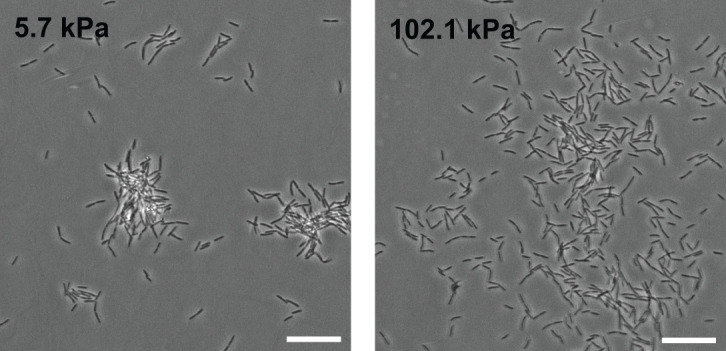
Phase contrast images of WT PAO1 bacteria on PEG hydrogels 5h after the onset of surface colonization. Scale bars, 20 μm.
Figure 1—figure supplement 3. Substrate rigidity impacts early microcolony morphology in the absence of shear flow.

Phase contrast images of WT PAO1 bacteria on PAA hydrogels at the bottom of a 6-well plate, after 150 min of incubation at 37°C without agitation. In red: initial adhering bacteria at t0. (Scale bars, 20 μm.).
Figure 1—figure supplement 4. In the T4P-deficient mutant PAO1 , substrate rigidity does not significantly impact colony morphology.

Colonies imaged after 5 h. Scale bar 20 μm.