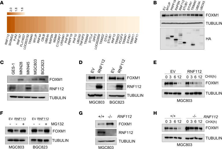
Figure 1. Screen and identification of an E3 ligase targeting FOXM1.
(A) Heatmap of the fold change in endogenous FOXM1 protein expression after the transfection with siRNA targeting E3 ligases in HEK293T cells. (B) Immunoblot detection of FOXM1 in HEK293T cells after transfection with the indicated plasmids. (C) Immunoblot analysis of endogenous expression of FOXM1 and RNF112 in GES-1 cells and a panel of human gastric cancer cell lines. (D) Immunoblot analysis of endogenous FOXM1 in MGC803 and BGC823 cells after transfection with RNF112-HA plasmids and EV for 48 hours. (E) Immunoblot analysis of endogenous FOXM1 after ectopic RNF112 expression in MGC803 cells in the presence of CHX (100 μg/mL) for the indicated time. (F) Immunoblot analysis of endogenous FOXM1 expression after ectopic RNF112 expression in MGC803 and BGC823 cells treated with 10 μM MG132 for 8 hours. (G) FOXM1 and RNF112 were detected by immunoblotting in WT and RNF112-depleted MGC803 cells. (H) Immunoblot analysis of endogenous FOXM1 in WT and RNF112-depleted MGC803 cells in the presence of CHX (100 μg/mL) for the indicated time. Complete unedited blots are listed in the supplemental material.

