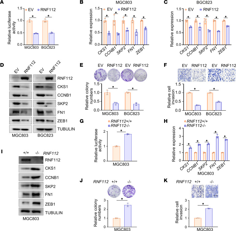
Figure 2. RNF112 suppresses FOXM1 transcriptional targets and inhibits the malignancy of gastric cancer cells in vitro.
(A) The luciferase activity of 6×FKH luciferase reporter after RNF112 overexpression. MGC803 and BGC823 cells in 24-well plates were transfected with 6×FKH luciferase reporter and pRL-TK, together with RNF112 plasmids or EV, respectively. The luciferase activity was measured 24 hours later (n = 3). (B–D) Quantitative reverse transcription–PCR (qRT–PCR) analysis (B and C) and immunoblot analysis (D) of the expression of FOXM1 target genes after transfection with RNF112-HA plasmids and EV for 48 hours in MGC803 and BGC823 cells (n = 3). (E and F) Representative images of colony-formation assays (E) and Transwell invasion assays (F) using stably overexpressing EV or RNF112 MGC803 and BGC823 cells. The relative number of colonies and invasive cells was normalized and plotted (n = 3). Scale bar: 400 μm. (G) The luciferase activity of 6×FKH luciferase reporter in WT and RNF112-depleted MGC803 cells (n = 3). (H and I) qRT–PCR analysis (H) and immunoblot analysis (I) of the expression of FOXM1 target genes in WT and RNF112-depleted MGC803 cells (n = 3). (J and K) Representative images of colony-formation assays (J) and Transwell invasion assays (K) using WT and RNF112-depleted MGC803 cells (n = 3). Scale bar: 400 μm. Data are presented as mean ± SD. Statistical significance was calculated using Student’s t test (A–C, E–H, J, and K). *P < 0.05. Complete unedited blots are in the supplemental material.

