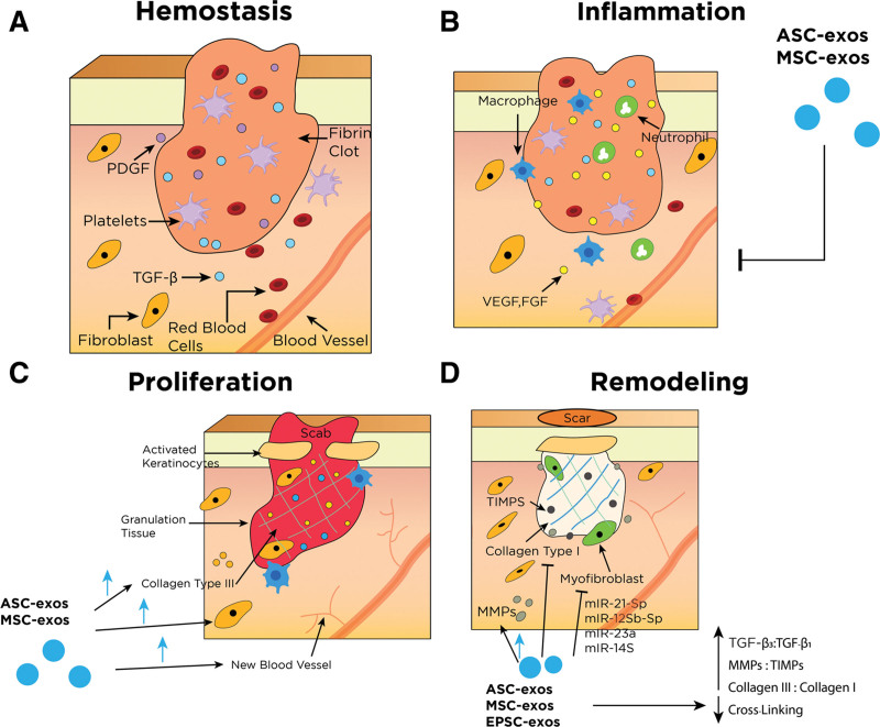
Fig. 4.
Simplified wound healing process and its main effectors. *Other key components have been removed for simplification. Refer to table, Supplementary Digital Content 2 for more details regarding different types of exosomes and their respective effects, http://links.lww.com/PRSGO/C605. A, Hemostasis begins immediately following a disruption of epithelial integrity. This phase is characterized by the restriction of blood flow and the formation of blood clots to the injury site to control bleeding. The clot and surrounding tissue release several cytokines and growth factors that recruit inflammatory cells (chemotaxis) to the injury site and progress to the next phase. B, Inflammatory phase is characterized by localized tissue swelling via influx of inflammatory cells (neutrophils and macrophages) and transudate. Inflammatory cells are recruited to the area to degrade cellular debris and prevent infection. Macrophages play an essential role in this phase as they promote the transition to the proliferative healing phase by stimulating keratinocytes, fibroblasts, and angiogenesis. Exosomes are thought to be able to tone down the inflammatory response and prevent secondary injury in this phase. C, Proliferative phase is characterized by reepithelialization. Fibroblasts lay down a scaffold of collagen III and ECM in the wound bed and strengthen new granulation tissue. Keratinocytes migrate across the wound surface to close the defect. Exosomes increase proliferation and migration of fibroblasts, collagen III deposition, and angiogenesis. D, Remodeling phase is the final phase of wound healing and can last for years. This stage is characterized by ECM and tissue remodeling. Collagen I begins to replace collagen III and cross-linking matures, leading to flattening of scars. Myofibroblasts contract and approximate wound edges. Exosomes reduce excessive cross-linking and increased ratios of collagen III: collagen I, MMPs:TIMPs, and TGF-β3:TGF-β1. Exosomes also inhibit myofibroblast differentiation via several miRNAs’ actions. ASC-exos indicates adipose-derived exosomes; MSC-exos, mesenchymal stem cell-derived exosomes; EPSC-exos, epidermal stem cell-derived exosomes; PDGF, platelet-derived growth factor; VEGF, vascular endothelial growth factor; EGF, epidermal growth factor; FGF, fibroblast growth factor; KGF, keratinocyte growth factor.

