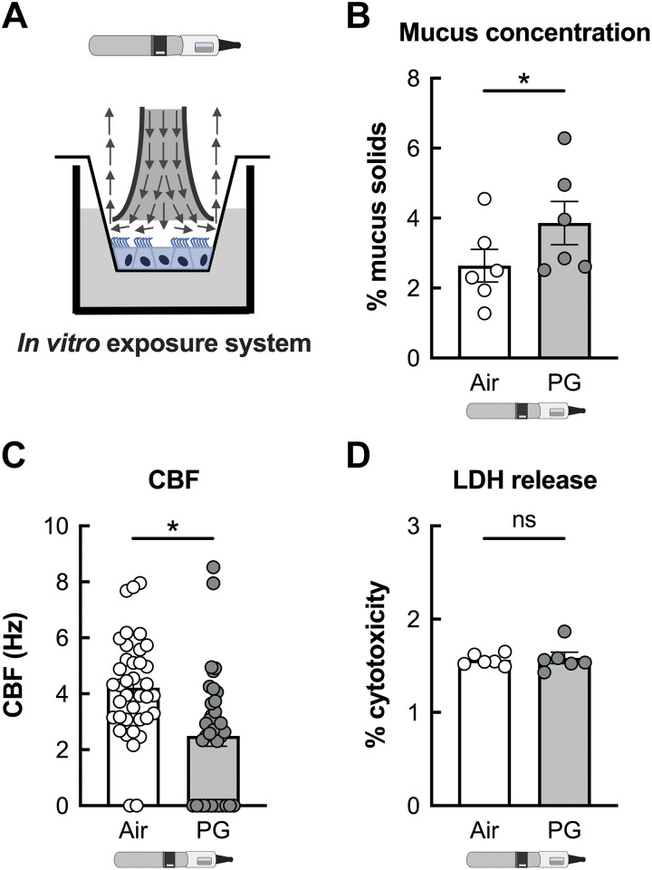
Figure 3.
PG e-cig aerosols induce mucociliary dysfunction in primary HBECs in vitro. A: representative schema illustrating exposure of fully differentiated primary HBECs to e-cig aerosols. Arrows indicate direction of aerosol flow. B: mucus concentrations are significantly increased in HBECs exposed to PG e-cig aerosols after 5 days compared with air-exposed controls. n = 6 from 5 lungs. C: ciliary beat frequency (CBF) of HBECs exposed to PG e-cig aerosols is significantly reduced after 5 days compared with air-exposed controls. n ≥ 36 from 4 lungs. D: 5-day exposure of HBECs to PG e-cig aerosols does not affect cytotoxicity as assessed by lactate dehydrogenase (LDH) release into basolateral media. n = 6 lungs. Data are presented as means ± SE. *P < 0.05, ns = not significant. Data were analyzed by two-tailed t test (B and D) or mixed-effects model (C) after assessing normality by Shapiro–Wilk. HBECs, human bronchial epithelial cells; PG, propylene glycol.

