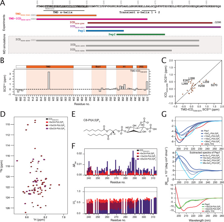
Figure 2. The intracellular juxtamembrane (ICJM) region of the prolactin receptor (PRLR) interacts with PI(4,5)P2.
(A) Overview of investigated PRLR variants. (B) Secondary chemical shifts (SCSs) of transmembrane domain (TMD)-intracellular domain (ICD)F206-S270 reconstituted in 1,2-dihexanoyl-sn-glycero-3-phosphocholine (DHPC) micelles. (C) Correlation plot of the SCSs of ICDG236-Q396 plotted against those of TMD-ICDF206-S270. (D) 15N,1H-HSQC spectra of 15N-ICDK235-G313 titrated with 5×, 10×, and 25× molar excess of C8-PI(4,5)P2. (E) Structure of C8-PI(4,5)P2. (F) Backbone amide chemical shift perturbations (CSPs) and peak intensity changes upon addition of C8-PI(4,5)P2 to 15N-ICDK235-G313 plotted against residue number. (G) Top: Far-UV CD spectra of Pep1 titrated with C8-PI(4,5)P2 or in 65% trifluorethanol (TFE). Middle: Far-UV CD spectra of Pep1 in the presence of 5 x-38x C8-PI(4,5)P2 subtracted with the spectrum of Pep1 in the absence of C8-PI(4,5)P2. Bottom: Far-UV CD spectra of Pep2 titrated with C8-PI(4,5)P2 or in 65% TFE.


