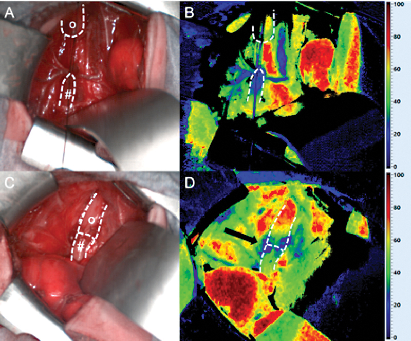
Fig. 1.

Intraoperative situs ( A, C ) and HSI StO 2 ( B, D ) after TEF ligation: Well-perfused upper esophageal pouch ( o ) and impaired perfusion of the lower esophagus ( # ) before resection of its distal tip ( A, B ). After resection of the tip of the distal pouch (Fig. 2), improved perfusion with only minor impairment at the anastomotic suture line itself ( arrow ) was detected ( C, D ). HSI, hyperspectral imaging; StO 2 , tissue oxygen saturation; TEF, tracheoesophageal fistula.
