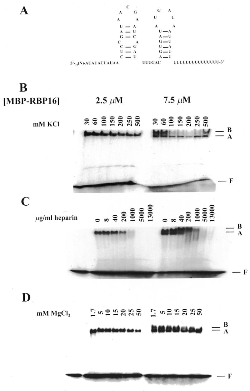
Figure 1.

Binding properties of RBP16. (A) Nucleotide sequence (41) and experimentally determined secondary structure (24) of gA6[14]. Note that this structure was not verified in our experiments. [32P]-labeled gA6[14] (10 fmol) was incubated with 2.5 and 7.5 µM RBP16 in the presence of increasing amounts of (B) KCl, (C) heparin and (D) MgCl2. The resulting protein–gRNA complexes were separated on a native 4% polyacrylamide gel in 50 mM Tris–glycine (pH 8.8). The position of the faster and slower migrating complexes is indicated by A and B, respectively. F represents the free gRNA.
