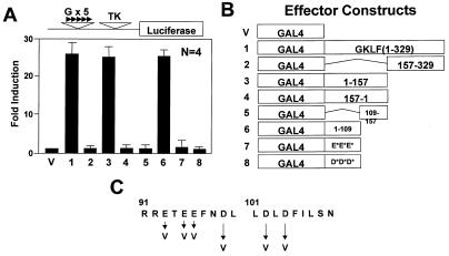
Figure 1.
Localization of the transactivation domain of GKLF by the GAL4 fusion assay. Chinese hamster ovary (CHO) cells were co-transfected with 5 µg/10 cm dish each of the indicated effector construct and the pG5TKLUC reporter. (A) Mean fold-induction in luciferase activity by the respective effector over that by the control vector containing only the DNA-binding domain of GAL4 (effector V). Bars represent standard deviations. (B) Schematic presentation of the various GAL4 fusion effector constructs. The point mutants involving the two clusters of acidic residues, effectors 7 and 8, are drawn as E*E*E* and D*D*D*, respectively. (C) Amino acid sequence between residues 91 and 110 of GKLF and identifies the mutagenized residues. All luciferase activities were standardized to the internal control β-galactosidase activities. Data represent the means of four independent experiments, each performed in duplicate.

