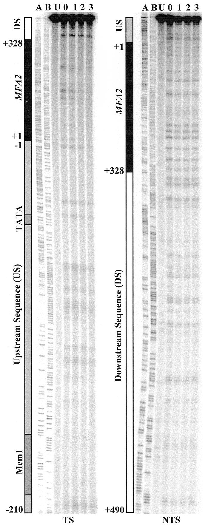
Figure 1.

Typical autoradiographs depicting UV-induced CPDs in the TS and NTS of a RAD26 wild-type strain. CPD repair was examined in the RsaI restriction fragment, which contains the MFA2 transcribed region (black bar), its upstream sequence (US, grey bar) and downstream sequence (DS, white bar). The Mcm1 binding site and the TATA box are indicated by dark grey shading in the control region. Numbers on the left of the bars denote the nucleotide position in relation to the MFA2 transcription start point. Ladder A, G+A; ladder B, C+T. Lane U, DNA from unirradiated cells; lane 0, DNA from cells receiving 125 J/m2 UV and extracted immediately; lanes 1–3, DNA from cells that received UV but which were incubated afterwards in medium for these times (h) before extraction.
