Figure 8.

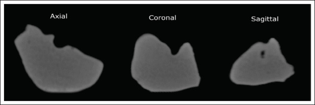
Appearance of the liver model in the CT scan in all three planes. Each view was sectioned approximately at the middle of the respective envelope dimension of the model.

Appearance of the liver model in the CT scan in all three planes. Each view was sectioned approximately at the middle of the respective envelope dimension of the model.