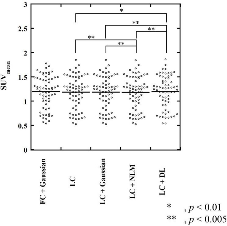
Figure 5.

SUVmean of background fibroglandular tissue. The SUVmean of the background fibroglandular tissue in the LC + DL images was slightly higher than that in the LC, LC + Gaussian, and LC + NLM images, whereas no significant difference in SUVmean was observed between the LC + DL and the reference (FC + Gaussian) images
