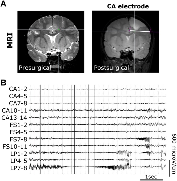
Figure 3.
Mutation-positive electrode (CA) in patient SE3. (A) Pre- and postsurgical 3T MRI of patient SE3, showing the trajectory of the mutation-positive electrode (CA) at the bottom of the dysplastic region not resected during the first surgery (white dots). (B) Representative SEEG traces of the contacts within the epileptogenic zone network from LP and FS mutation-negative electrodes and the CA mutation-positive electrode in the propagation zone network. CA, anterior cingular; FS, superior frontal; LP, posterior limit.

