Figure 4.
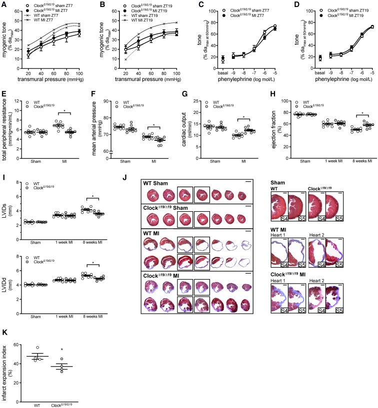
Global ClockΔ19/Δ19 mutation attenuates myogenic responsiveness and improves cardiac function following myocardial infarction. Myogenic tone at (A) ZT7 (n = 7–8) and (B) ZT19 (n = 7–8) in ClockΔ19/Δ19 cremaster arteries isolated from sham-operated mice and mice with a myocardial infarction (MI). Traces from sham and MI wild-type (WT) mice are reproduced from Figure 3A for qualitative comparison purposes. Phenylephrine-stimulated vasoconstriction at (C) ZT7 (n = 7–8) and (D) ZT19 (n = 7–8) in ClockΔ19/Δ19 cremaster arteries isolated from sham-operated and MI mice. Pressure–volume hemodynamic assessments of (E) total peripheral resistance, (F) mean arterial pressure, and (G) cardiac output in WT and ClockΔ19/Δ19 mutant mice with sham or MI procedure (n = 8). Echocardiographic assessments of (H) ejection fraction and (I) systolic and diastolic left ventricular internal diameter (LVIDs and LVIDd) in WT and ClockΔ19/Δ19 mutant mice with sham or MI procedure (n = 8). (J) Cardiac histology was processed at 8 weeks post-MI. Shown are representative histological images of cardiac infarcts in WT and ClockΔ19/Δ19 mutant mice with sham or MI procedure; serial sections are shown on the left (bar = 2 mm) and close-ups of the infarct region in serial sections 4–5 are shown on the right (bar = 500 µm). (K) Quantification of infarct expansion from the histological images (n = 4). All data are statistically analysed as unpaired single comparisons using t-tests or Mann–Whitney tests, with * denoting P < 0.05.

