
Figure 5.
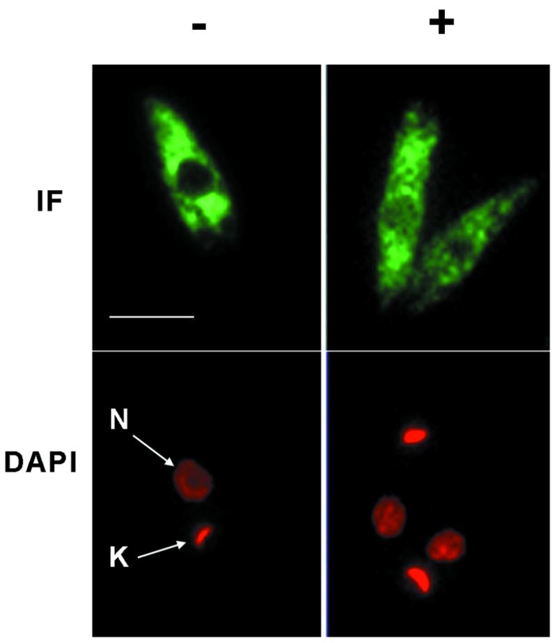
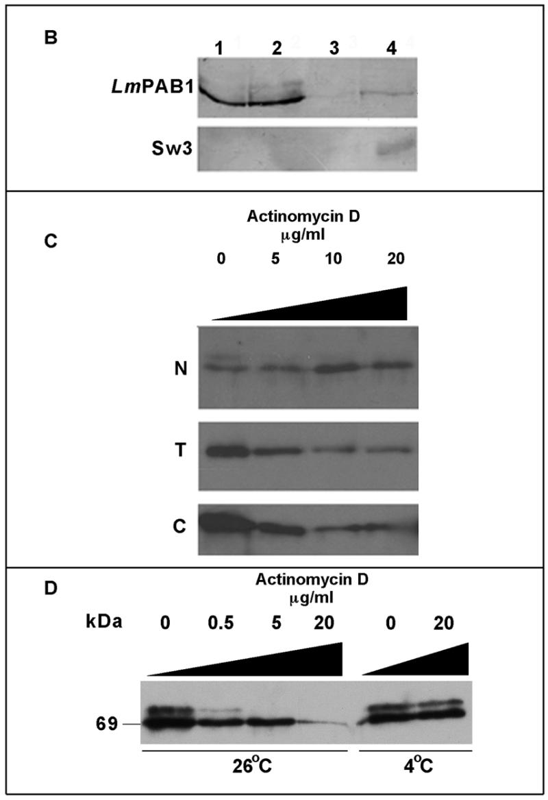
Transcriptional inhibition affects the cellular localisation of LmPAB1. (A) Immunofluorescent detection of LmPAB1 (with abSK375; IF) and DAPI staining of nuclear (N) and kinetoplast (K) DNA in late logarithmic phase parasites, untreated (–) or incubated with 10 µg/ml actinomycin D (+) for 10 h prior to fixing and permeabilisation. Scale bar = 5 µm. (B) Parasite fractions were separated (in the presence of protease inhibitors) and immunoblotted with anti-LmPAB1 (8% SDS–PAGE) or ab415 (Sw3, anti-histone H1; 10% SDS–PAGE). Lane 1, total lysate, 5 × 10⁶ cells; lane 2, cytoplasmic fraction, 5 × 10⁶ cells; lane 3, nuclear fraction, 5 × 10⁶ cells; lane 4, nuclear fraction, 3 × 10⁷ cells. (C) Parasites treated with 0–20 µg/ml of actinomycin D for 10 h were fractionated (in the absence of protease inhibitors) and analysed by 6% SDS–PAGE and immunoblotting with anti-LmPAB1. Chemiluminescent exposures were for 15 s [total (T) and cytoplasmic (C) fractions from 2.5 × 10⁷ cells] or 1 min [nuclear fraction (N) from 4.5 × 10⁷ cells]. (D) Cytoplasmic fractions from 2 × 10⁶ parasites incubated with actinomycin D for 24 h at 26°C or 4°C as indicated. LmPAB1 was immunodetected by chemiluminescence, exposure time 8 min.