Fig. 1.

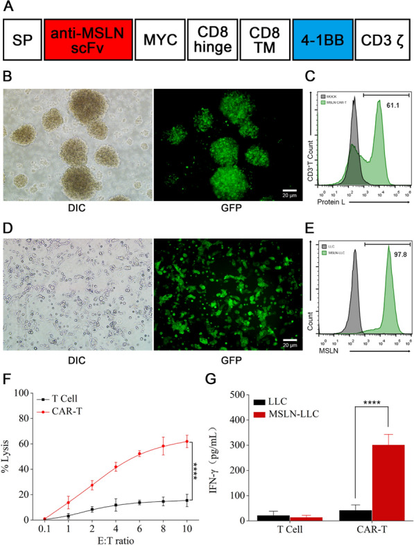
Preparation and characterization of CAR-T cells. A Molecular design of anti-MSLN mCAR. B The transfection efficiency of lentiviral in T cells was assessed by GFP expression using fluorescence microscopy. Scale bar: 20 μm. C Anti-MSLN CAR expression was assessed by Protein L staining and flow cytometry. Representative Protein L staining results are shown for mock transduced T cells and MSLN CAR-T cells. D Fluorescence images of LLC cells stably expressing GFP-MSLN after puromycin selection. Scale bar: 20 μm. E Representative flow cytometry histogram of the expression level of MSLN on LLC cells. An anti-MSLN antibody was used to confirm MSLN expression. F Cytolysis of CAR-T cells against MSLN-LLC cells. The cytotoxic activity of CAR-T and control T cells against cancer cell lines was assessed by an LDH-release assay at the indicated effector-to-target (E:T) ratios. G The level of IFN-γ evaluated by ELISA after co-culture of CAR-T cells with MSLN-LLC cells for 24 h. Data are presented as the mean ± SD of three biological replicates. *P < 0.05, **P < 0.01, ***P < 0.001, ****P < 0.0001, ns: not significant
