Fig. 3.
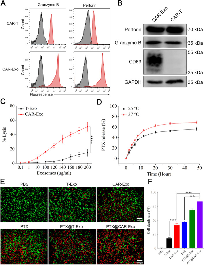
The enhanced antitumor effect of PTX@CAR-Exos in vitro. A Flow cytometric analyses of granzyme B and perforin expression of CAR exosomes conjugated to latex beads or CAR-T cells. The histograms shown isotype controls (black) and positive expression (red). B The expression of granzyme B and perforin in CAR-Exos or whole-cell lysates of CAR-T cells were analyzed by western blotting. C The cytotoxic activity of different concentrations of CAR exosomes against MSLN-LLC cells. D In vitro drug release profile of paclitaxel-loaded CAR exosomes was evaluated in PBS at pH 7.4 and different temperatures. The percentage of drug release (%) = OD value of the PTX released from the CAR exosomes /OD value of the total PTX in the CAR Exosomes × 100%. E Cytotoxicity and Calcein-AM/PI staining analysis in vivo. Fluorescent microscopic imaging of MSLN-LLC cells after treatment with PBS, T-Exos, CAR-Exos, PTX, PTX@T-Exos, or PTX@CAR-Exos for 24 h, cells were stained with Calcein-AM/PI (original magnification, 10 ×). Scale bar: 100 μm. F Quantitative analysis of cell death rate analyzed by Calcein-AM/PI staining. Data are presented as the mean ± SD of three biological replicates. *P < 0.05, **P < 0.01, ***P < 0.001, ****P < 0.0001, ns: not significant

