. 2023 Jun 1;43(6):e220177. doi: 10.1148/rg.220177

Figure 2.

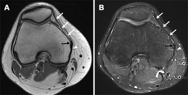
MPFL. Axial proton-density–weighted (A) and T2-weighted fat-suppressed (B) MR images show a normal MPFL (white arrows). The MPFL is a horizontally oriented ribbonlike ligament that spans from the medial patella to the adjacent medial femur. The MPFL merges with the fibers of the medial collateral ligament (black arrow) at the epicondyle and inserts immediately posterior onto the femur (arrowhead).

MPFL. Axial proton-density–weighted (A) and T2-weighted fat-suppressed (B) MR images show a normal MPFL (white arrows). The MPFL is a horizontally oriented ribbonlike ligament that spans from the medial patella to the adjacent medial femur. The MPFL merges with the fibers of the medial collateral ligament (black arrow) at the epicondyle and inserts immediately posterior onto the femur (arrowhead).