. 2023 Jun 1;43(6):e220177. doi: 10.1148/rg.220177

Figure 20.

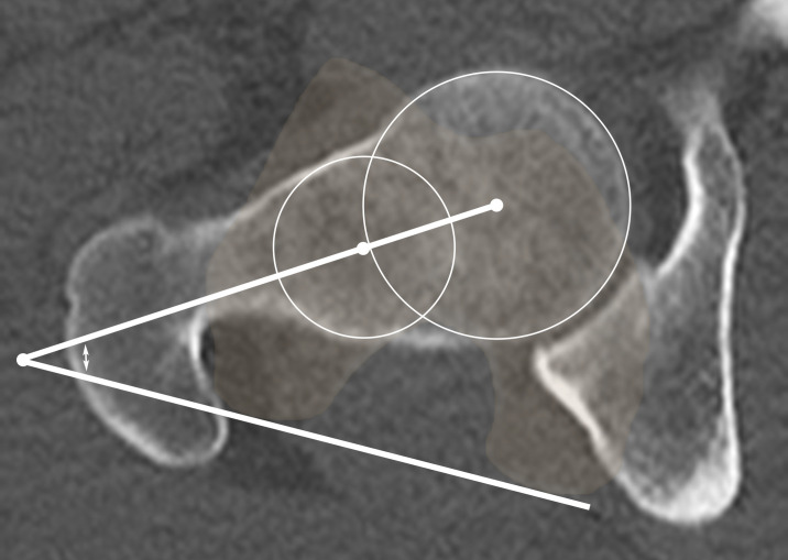
Measurement of femoral version. Axial CT image shows the femoral neck angle (double-headed arrow) measurement obtained by using 3D best-fit circles at the femoral head and femoral neck. The femoral version is determined by combining this measurement with the femoral bicondylar axis at the knee. Using 3D techniques for measurements can limit inter- and intraobserver error and improve accuracy.

Measurement of femoral version. Axial CT image shows the femoral neck angle (double-headed arrow) measurement obtained by using 3D best-fit circles at the femoral head and femoral neck. The femoral version is determined by combining this measurement with the femoral bicondylar axis at the knee. Using 3D techniques for measurements can limit inter- and intraobserver error and improve accuracy.