. 2023 Jun 1;43(6):e220177. doi: 10.1148/rg.220177

Figure 26.

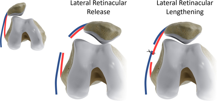
Lateral patellar retinacular release versus lengthening. Superficial (blue) and deep (red) layers of the lateral patellar retinaculum are schematically depicted. In the retinacular release procedure, which is frequently performed arthroscopically, both layers are transected in one plane. Full lateral release may cause iatrogenic medial patellar subluxation and instability. In retinacular lengthening, deep and superficial layers are isolated during an open surgery and transected at different levels to allow the patella to reduce and then are reapproximated to achieve appropriate tension and maintain integrity of the lateral retinaculum.

Lateral patellar retinacular release versus lengthening. Superficial (blue) and deep (red) layers of the lateral patellar retinaculum are schematically depicted. In the retinacular release procedure, which is frequently performed arthroscopically, both layers are transected in one plane. Full lateral release may cause iatrogenic medial patellar subluxation and instability. In retinacular lengthening, deep and superficial layers are isolated during an open surgery and transected at different levels to allow the patella to reduce and then are reapproximated to achieve appropriate tension and maintain integrity of the lateral retinaculum.