. 2023 Jun 1;43(6):e220177. doi: 10.1148/rg.220177

Figure 28.

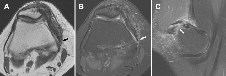
Recurrent patellar dislocation in a 29-year-old woman 3 years after she underwent MPFL reconstruction. Axial proton-density–weighted (A) and T2-weighted (B) fat-suppressed MR images and sagittal T2-weighted fat-suppressed MR image (C) show tearing of the femoral attachment site of the reconstructed MPFL (arrow), near the femoral interference screw.

Recurrent patellar dislocation in a 29-year-old woman 3 years after she underwent MPFL reconstruction. Axial proton-density–weighted (A) and T2-weighted (B) fat-suppressed MR images and sagittal T2-weighted fat-suppressed MR image (C) show tearing of the femoral attachment site of the reconstructed MPFL (arrow), near the femoral interference screw.