. 2023 Jun 1;43(6):e220177. doi: 10.1148/rg.220177

Figure 3.

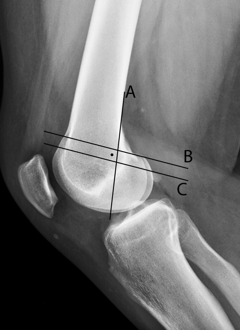
Radiographic landmarks for MPFL reconstruction. The Schöttle point (dot) is the midpoint of the MPFL attachment on the femur and the target for MPFL reconstruction tunnel placement (16). Lateral radiograph shows that the Schöttle point is 1 mm anterior to the posterior cortex extension line (A), distal to a line drawn between the anterior and posterior origins of the medial femoral condyle (B), and proximal to the level of the posterior point of the Blumensaat line (C).

Radiographic landmarks for MPFL reconstruction. The Schöttle point (dot) is the midpoint of the MPFL attachment on the femur and the target for MPFL reconstruction tunnel placement (16). Lateral radiograph shows that the Schöttle point is 1 mm anterior to the posterior cortex extension line (A), distal to a line drawn between the anterior and posterior origins of the medial femoral condyle (B), and proximal to the level of the posterior point of the Blumensaat line (C).