. 2023 Jun 1;43(6):e220177. doi: 10.1148/rg.220177

Figure 8.

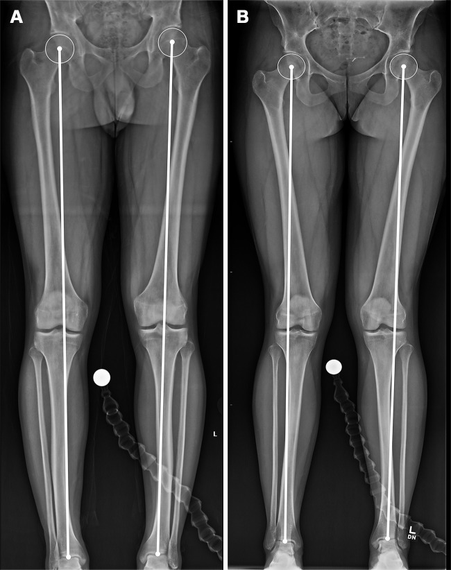
Evaluation of the mechanical axis on radiographs in a 27-year-old man with a normal mechanical axis (A) and a 31-year-old woman with bilateral genu valgum (B). The mechanical axis is evaluated on standing full-length lower extremity radiographs. A line is drawn from the center of the femoral head to the center of the talar dome. A normal mechanical axis is defined by a line passing through the knee between the medial and lateral margins of the tibial spine.

Evaluation of the mechanical axis on radiographs in a 27-year-old man with a normal mechanical axis (A) and a 31-year-old woman with bilateral genu valgum (B). The mechanical axis is evaluated on standing full-length lower extremity radiographs. A line is drawn from the center of the femoral head to the center of the talar dome. A normal mechanical axis is defined by a line passing through the knee between the medial and lateral margins of the tibial spine.