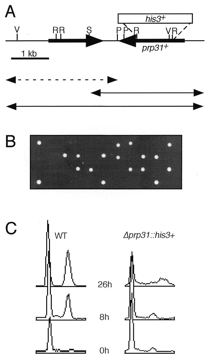
Figure 3.

Analysis of the prp31+ locus and disruption phenotype. (A) Schematic of the genomic clone containing the prp31+ gene. Double-headed arrows represent subclones that were transformed into the prp31-E1 mutant strain FY1138 to test for complementation activity scored by the ability to form colonies at 36°C. Continuous lines, subclones that were able to complement at 36°C; broken line, subclone that failed to complement at 36°C. The box above the line indicates the location of the disruption/deletion with the his3+ marker. V, EcoRV; R, EcoRI; S, SphI; P, SpeI. (B) Tetrads dissected from the diploid prp31::his3+/prp31+ were grown on YES at 32°C for 2 days. All growing spore clones were His– (data not shown). (C) Flow cytometry of germinating wild-type (WT) and Δprp31::his3+. Samples were taken at 0, 8 and 26 h from cells growing in selective medium.
