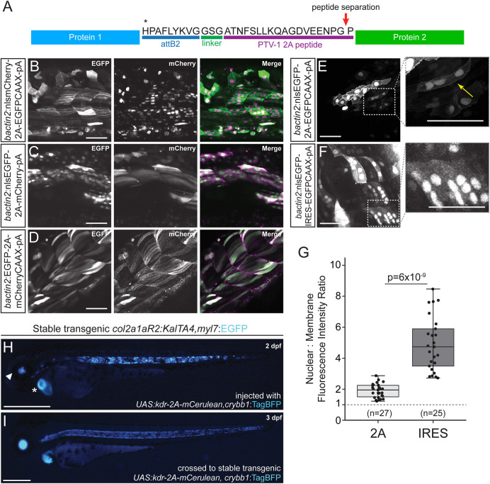
Fig. 4.
2A peptide linkers for fluorescent protein tagging. (A) Schematic of protein product resulting from a Tol2 construct containing two protein sequences separated by a 2A peptide following the attB2 site of a Multisite Gateway expression construct. Translation of the first protein occurs, the first polypeptide is released at the site of peptide separation and translation of the second ORF proceeds with the same ribosome, resulting in equimolar protein amounts. (B-F) Transient injections of bactin2 promoter-driven fluorophore combinations separated by a 2A peptide tag. nlsmCherry-2A-EGFPCAAX (B), nlsEGFP-2A-mCherry (C) and EGFP-2A-mCherryCAAX (D). Transient injection of bactin2:nlsEGFP-2A-EGFPCAAX (E) demonstrates expression of EGFP in both the nucleus and membrane of cells, whereas injection of bactin2:nlsEGFP-IRES-EGFPCAAX (F) results in poor EGFP-CAAX detection in cells that are positive for nuclear EGFP. (G) Ratio of nuclear to membrane fluorescence intensity with n=total number of cells assayed (numbers at the base of the graph); for each condition, cells from five different embryos were analyzed. Welch's t-test (unequal variance); first and third quartiles are boxed, bars extend to the highest value within the 1.5× inter-quartile range. (H,I) Use of 2A fusions in Gal4/UAS transgene experiments. Injection of UAS:kdr-2A-mCerulean,crybb1:TagBFP into stable transgenic col2a1aR2:KalTA4,myl7:EGFP that expresses codon-optimized Gal4 in the developing notochord and subsequent cartilage lineages shows mosaic mCerulean expression in the developing notochord (H), with the secondary myl7:EGFP transgenic marker indicated with an asterisk and the crybb1:TagBFP marker indicated with an arrowhead. Homogenous notochord expression of mCerulean in stable F2 UAS:kdr-2A-mCerulean,crybb1:TagBFP crossed with col2a1aR2:KalTA4, myl7:EGFP (I). Scale bars: 50 μm (B-F); 500 μm (H,I).

