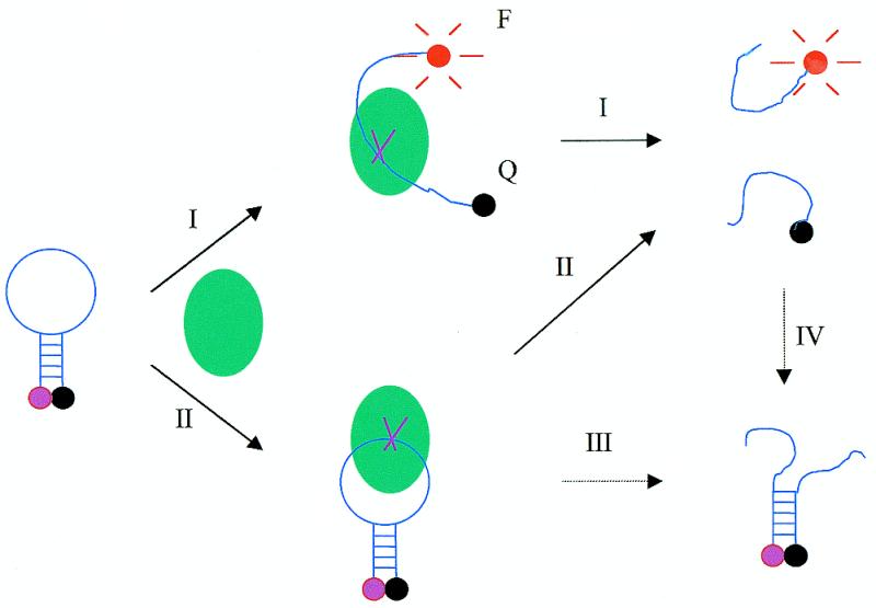
Figure 1.

Schematic representation of the fluorescence mechanism of the molecular beacon during cleavage by single-strand-specific DNA nuclease (indicated by solid arrows). The solid arrows indicate two paths (I and II) leading to fluorescence enhancement during digestion. The dashed arrows represent two possible processes (III and IV) in which no fluorescence enhancement is produced. Only the first cut is shown here. Even though the nuclease may keep on cutting one single strand many times, only the first cut contributes to the fluorescence signal increase. The ball represents the nuclease. MB, F and Q represent molecular beacon, fluorophore and quencher, respectively. Here the fluorophore and quencher are tetramethylrhodamine (TAMRA) and 4-(4′-dimethylaminophenylazo)benzoic acid (DABCYL), respectively.
