
Figure 4.
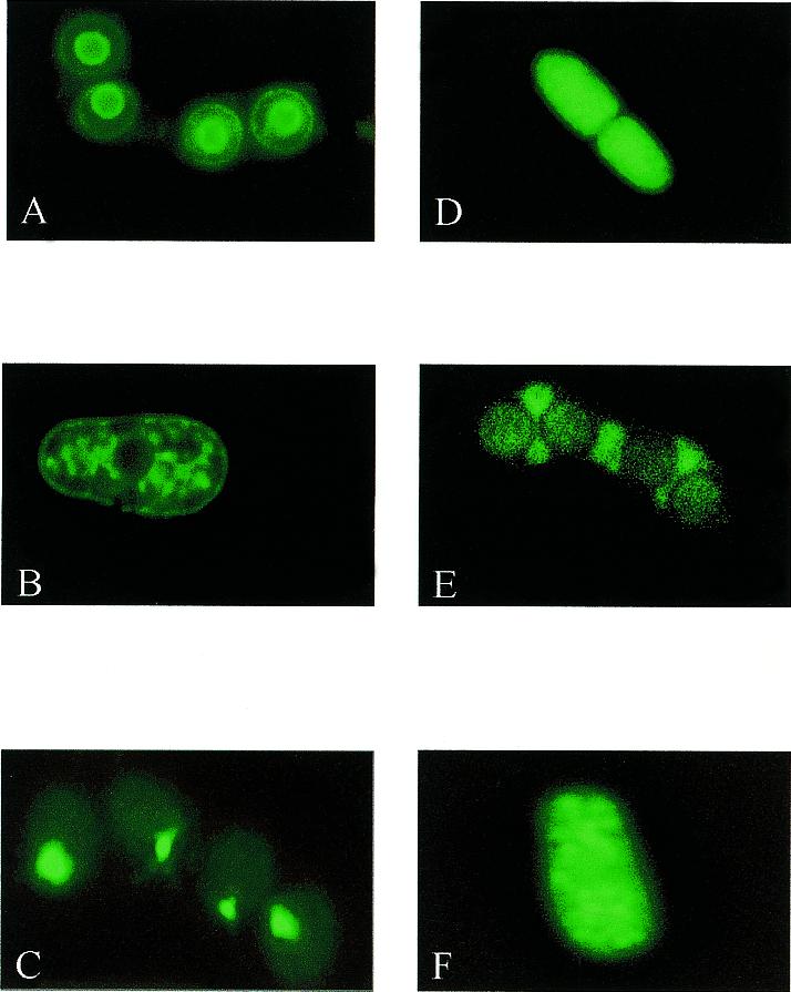
GFP fusion proteins displaying different cellular localizations. Living cells expressing GFP fusion proteins as genomic integrants were viewed by fluorescence microscopy. (A) lt13-26 (predominant staining in nuclear envelope and spore cytoplasm); (B) lt14-20 (nuclear envelope and ER); (C) lt1-4 (punctate staining proximal to nucleus in spores); (D) lt2-20 (cytoplasmic in septum-defective mutant); (E) lt3-2 (cytoplasmic in forming ascus); (F) lt2-3 (vacuolar). Images were taken with a Leica fluoresence microscope equipped with a high performance CCD camera (Sensicam) and Slidebook software (Intelligent Imaging Systems).