. 2023 Jun 15;24(6):510–523. doi: 10.1631/jzus.B2200447

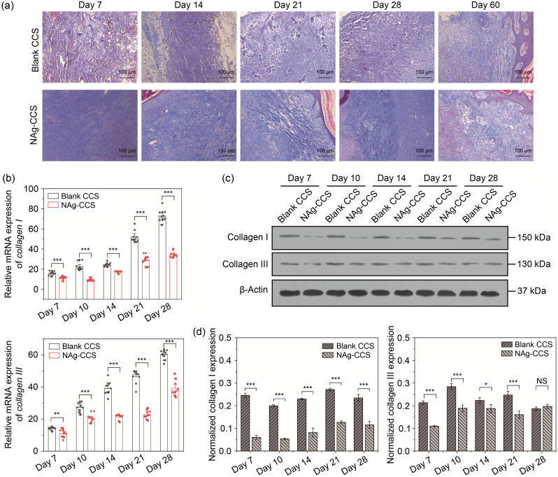
Fig. 5. Evaluation of collagen synthesis and deposition in miniature pigs following scaffold implantation. (a) Masson's trichrome staining of the back skin tissues of miniature pigs in the blank CCS (upper panels) and NAg-CCS (lower panels) groups after scaffold implantation on postoperative Days 7, 14, 21, 28, and 60. (b) The mRNA expression levels of collagen I (upper panel) and collagen III (lower panel) detected by qRT-PCR. (c) Representative images of western blotting showing the protein expression of collagen I and collagen III in the two groups at different postoperative time points. (d) Relative protein expression (western blotting) of collagen I and collagen III in different groups at different postoperative time points. Data are expressed as mean±standard deviation (SD) ( n=3). P<0.05, ⁎⁎ P<0.01, ⁎⁎⁎ P<0.001; NS: not significant. NAg: nanosilver; CCS: collagen-chitosan scaffold; mRNA: messenger RNA; qRT-PCR: quantitative real-time polymerase chain reaction.

Fig. 5